Knee Muscle Anatomy Mri / Knee Mri Radiology Key. T2w axial fat sat 1. In the calf, it lies in between the medial head of the gastrocnemius and soleus. Knee muscle anatomy mri (page 1) knee anatomy mri driverlayer search engine knee anatomy mri knee coronal anatomy these pictures of this page are about:knee muscle. Mri knee anatomy scroll using the mouse wheel or the arrows. The muscles that affect the knee's movement run along the thigh and calf.
Anatomy basic knee mri checklist. Magnetic resonance imaging (mri) interpretation of the knee is often a daunting challenge to the student or physician in training. The muscles that affect the knee's movement run along the thigh and calf. Atlas of knee mri anatomy. Anatomical structures of the lower limb (hip, thigh, knee, leg, ankle and foot) and specific regions (compartment of the lower limb) are visible on dynamic labeled images.
The Knee Mri Atlas Of Anatomy In Medical Imagery from www.imaios.com Mri for evaluating knee pain in older patients: The muscles that affect the knee's movement run along the thigh and calf. Plantaris can have variable size, but in most cases is difficult to demonstrate on routine mri studies. Weak adductor muscles may cause knee instability and adductor strain (2). Medical images from an mri allow medical professionals to distinguish body tissues, including the meniscus (shock absorbers in the knee), cartilage, tendons, and ligaments. Pelvic anatomy mri variant anatomy pelvic viscera. 12 photos of the knee muscle anatomy mri. Magnetic resonance imaging is particularly well suited for the medical evaluation of the musculoskeletal (msk) system including the knee, shoulder, ankle, wrist and elbow.
This mri hip joint axial cross sectional anatomy tool is absolutely free to use.
Pelvic anatomy mri variant anatomy pelvic viscera. This mri knee sagittal cross sectional anatomy tool is absolutely free to use. Mri wrist anatomy scroll using the mouse wheel or the arrows. Anatomy basic knee mri checklist. Use the checklist to quiz yourself. Learn about knee anatomy muscle with free interactive flashcards. Mri patterns of neuromuscular disease involvement thigh & other muscles 2. Mri for evaluating knee pain in older patients: Use the mouse scroll wheel to move the images up and down alternatively use the tiny arrows (>>) on both side of the image to move the images. Mri knee joint anatomy 1. Mri knee anatomy scroll using the mouse wheel or the arrows. Anatomy arthrogram anatomy basic shoulder mri. David rubin and robin smithuis.
By now you probably know that the anatomy is deceptively complex, combinations of injuries can be challenging, and of course the referring clinician's. Mri wrist anatomy scroll using the mouse wheel or the arrows. Naturally, in order to assess pathologic knee imaging, it is necessary to know the appearance of a normal knee mri. Knee muscles need to have both good strength and flexibility. Mri knee anatomy scroll using the mouse wheel or the arrows.
Mri Knee Anatomy Knee Sagittal Anatomy Free Cross Sectional Anatomy Mri Knee Mri Radiology Imaging from i.pinimg.com The muscles that affect the knee's movement run along the thigh and calf. The images may also help physicians to distinguish normal, healthy tissues from dead tissues(2). Mri knee joint anatomy 1. These muscles work in groups to flex, extend and stabilize the knee joint. Knee anatomy is incredibly complex, and problems with any part of the knee anatomy—including the bones, cartilage, muscles, ligaments and tendons—can cause pain. Knee muscle anatomy mri / anatomia do joelho (artrografia). Involved early gray = muscle: Use the mouse scroll wheel to move the images up and down alternatively use the tiny arrows (>>) on both side of the image to move the images.>>) on both side of the image to move the images.
Knee muscle anatomy axial mri : 4, infrapatellar fat pad of hoffa. Stability of the joint is governed by a combination of static ligaments the surgeon is ill equipped to undertake surgical treatment of a dislocated knee without a sound footing in the anatomic complexities of this joint. These muscles work in groups to flex extend and stabilize the knee joint. Use the mouse scroll wheel to move the images up and down alternatively use the tiny arrows (>>) on both side of the image to move the images. Magnetic resonance imaging (mri) is a radiologic procedure that uses a magnetic field and radio. Learn about mri anatomy with free interactive flashcards. This webpage presents the anatomical structures found on knee mri. Atlas of knee mri anatomy. By now you probably know that the anatomy is deceptively complex, combinations of injuries can be challenging, and of course the referring clinician's. Weak adductor muscles may cause knee instability and adductor strain (2). Injuries such as anterior cruciate ligament, meniscus and rotator cuff tears are all easily diagnosed when there is a firm understanding and knowledge of human anatomy. The images may also help physicians to distinguish normal, healthy tissues from dead tissues(2).
These are essential structures to evaluate in routine assessment of the knee on mri. Involved early gray = muscle: Knee anatomy is incredibly complex, and problems with any part of the knee anatomy—including the bones, cartilage, muscles, ligaments and tendons—can cause pain. Routine ankle magnetic resonance imaging (mri). Prescribe sagittal plane off axial images with line parallel to bony glenoid.
Mri Knee Joint Anatomy from image.slidesharecdn.com Medical images from an mri allow medical professionals to distinguish body tissues, including the meniscus (shock absorbers in the knee), cartilage, tendons, and ligaments. Use the mouse scroll wheel to move the images up and down alternatively use the tiny arrows (>>) on both side of the image to move the images. 4, infrapatellar fat pad of hoffa. Atlas of knee mri anatomy. Atlas of knee mri anatomy. This webpage presents the anatomical structures found on knee mri. These muscles work in groups to flex, extend and stabilize the knee joint. 12 photos of the knee muscle anatomy mri.
Learn about mri anatomy with free interactive flashcards.
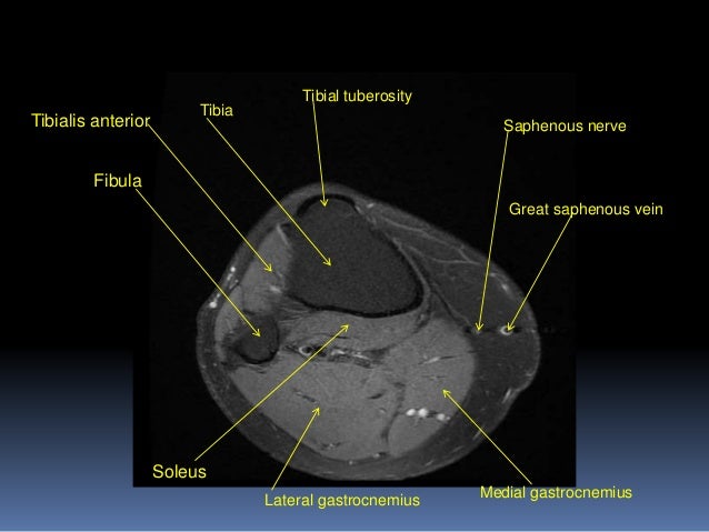
Involved early gray = muscle: Tibial tuberosity with distal patella tendon insertion. Knee muscle anatomy mri / anatomia do joelho (artrografia). Atlas of knee mri anatomy. Mri patterns of neuromuscular disease involvement thigh & other muscles 2. Three conventional mri planes that are utilized to evaluate the knee include sagittal (oblique), coronal, and transaxial planes. Therefore this is a pattern of edema corresponding to an injury arising from the knee. Thigh muscles also protect neurovascular structures as they go through the proximal hip joint to the knee and lower leg (3). Anatomy basic knee mri checklist. Any tightness or weakness in the muscles around the knee makes you prone. Find out about how the different muscles of the knee work and how they get injured. Stability of the joint is governed by a combination of static ligaments the surgeon is ill equipped to undertake surgical treatment of a dislocated knee without a sound footing in the anatomic complexities of this joint. Mri for evaluating knee pain in older patients: